Research highlight
Speeding up MRI: shorter scan times to increase availability
After real-world testing in an outpatient cognitive service, a new protocol shows how an accelerated procedure can cut scan time without compromising diagnostic utility.
Funding from Alzheimer’s Society has helped develop and test a highly accelerated MRI protocol. This protocol cuts total scan time to just under 7 minutes from 20 minutes or more, achieving substantial time savings without impacting reliability of a neuroradiologists assessment and could increase access to MRI while reducing costs of scans and reducing burden for patients.
With increasing numbers of disease modifying treatments being explored, ensuring MRI imaging is accessible to all is a priority. Especially given to access disease modifying treatments, people living with dementia require MRI scans as part of their routine investigations.
Dr Miguel Rosa-Grilo, Professor Nick Fox, Professor Geoff Parker and colleagues performed the first explorative study to understand how innovations in parallel imaging could be used in clinics to accelerate MRI imaging moving towards a reality where every person living with dementia could be diagnosed with a scan.
Indicators before dementia symptoms
The project builds on a history of highly influential Alzheimer’s Society-funded research. In the early 1990s, we funded Professor Nick Fox – then a research fellow – to investigate if early indicators of Alzheimer’s disease could be seen in the brains of people at risk before symptoms appeared.
From this funding, Nick published the first evidence that MRI scanning could measure cerebral volume change very precisely – and detect Alzheimer’s disease much earlier, even presymptomatically.
While MRI imaging was first used to produce a brain scan in the 1970s, and cerebral atrophy was a known characteristic of Alzheimer’s disease, the large variation between individuals made this difficult to use as a diagnostic technique.
Working with Professor Martin Rossor, Nick developed a registration-based method that determines how the boundaries of the scanned brain have moved and the resulting volume change – or how the brain is shrinking. They went on to validate this method, termed the Boundary Shift Integral technique, which became the industry standard for measuring atrophy progression in clinical trials.
Making scans accessible to all
Fast forward 50 years and structural imaging, such as CT and MRI scans, is recommended in the suite of tests as part of dementia diagnosis, to first rule out any other – potentially treatable – causes of cognitive decline and to assist with dementia subtype diagnosis.
Ideally, this would mean an MRI scan as it enables easier differentiation between different dementia subtypes and avoids exposing patients to radiation. However, there is a shortage of MRI scanners, and these scans take longer and are more costly than CT scans.
It was this problem that Professor Geoff Parker proposed addressing in his latest Alzheimer’s Society Project, for which we awarded a grant in 2023 supported by Alzheimer’s Society’s Heather Corrie Impact Fund.
Our research has taken advantage of recent breakthroughs in scanner technology. Our task was to work out just how fast we could scan while maintaining image quality good enough for diagnosis.
– Professor Geoff Parker, University College London
Reducing scan time by 63%
Using an ultra-fast MRI scan, the method speeds up the acquisition time by taking advantage of faster software pipelines that require no hardware upgrades to the scanner. If needed, machine learning techniques can be subsequently applied to further enhance the quality of the scan.
Geoff and colleagues have now published the first of the results from this study, comparing the ultra-fast method against the standard clinical scan.
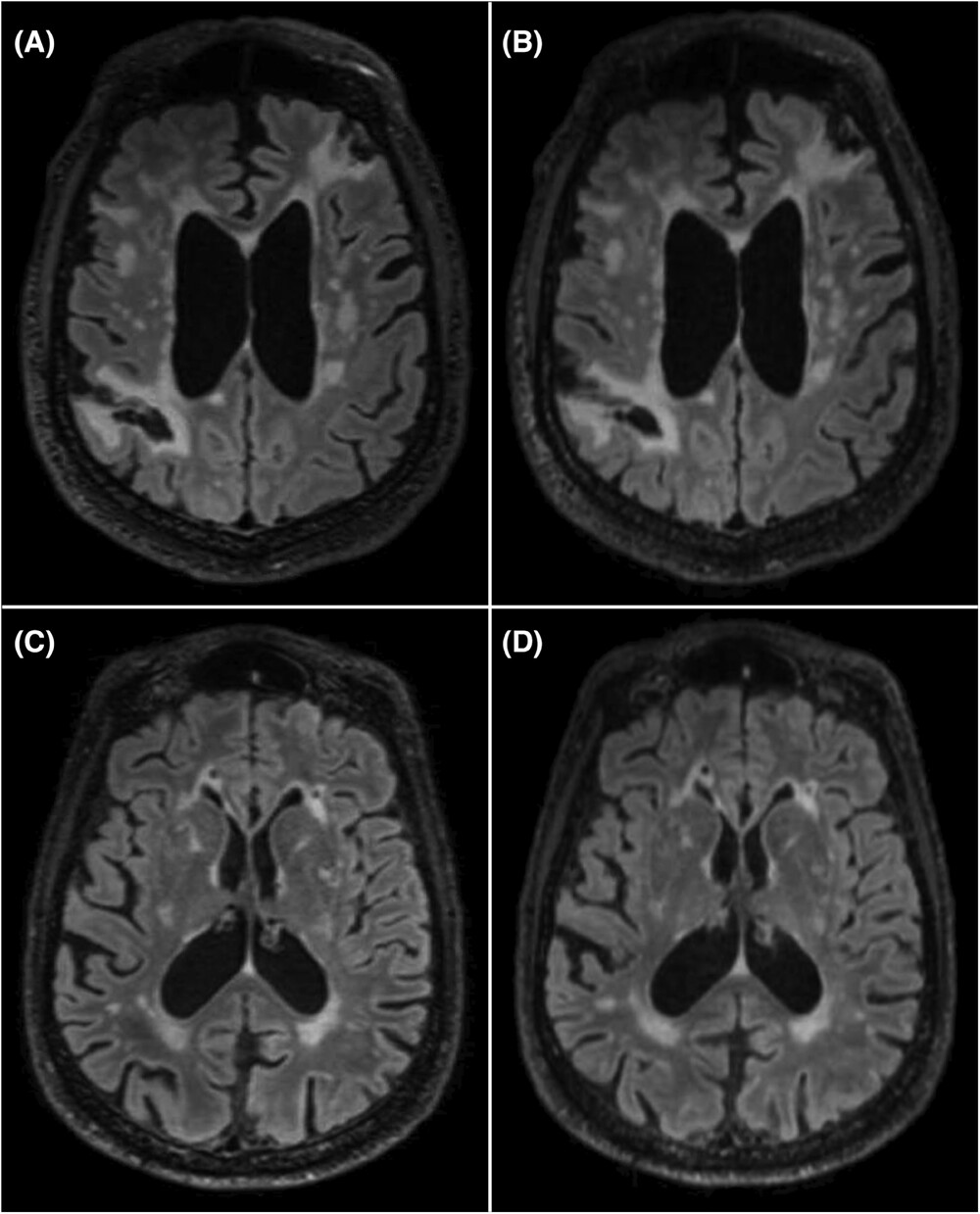
Examples of MRI sequences from the clinical (A, C) and fast (B, D) protocols. Figure from Rosa-Grilo M et al. (2025). doi: 10.1002/alz.70341
Overall, total acquisition (scan) time was markedly reduced, by 63%, so the scan could be performed in one third of the time taken previously. Reliability of the neurologist’s assessments of the newly accelerated protocol was not inferior to the standard of care protocol.
We were confident that the new scan would prove non-inferior to the standard scan, given the high image quality – but it was remarkable how well it performed.
– Dr Miguel Rosa-Grilo, University College London
This latest study from Miguel, Geoff and Nick was performed using a real-world approach, increasing the generalisability of results. People were invited to join the study through an outpatient service where an MRI brain scan had been planned as part of their routine assessment and the new scans followed typical workflows already used in the clinic. Over 95% of people approached to take part in the study, accepted.
MRI urgency: disease modifying treatments
Knowing dementia impacts the brain before symptoms means we can try to treat these conditions to prevent or slow the onset of dementia, which impacts quality of life.
Emerging disease modifying treatments, such as lecanemab and donanemab, require an MRI scan before starting treatment as well as for safety monitoring during treatment. With treatment ongoing for many months, up to 30 in some cases, this is not an insignificant number of scans.
This creates an urgent need to find ways to enable MRI scans to be accessible, by making them faster and cheaper. A shorter time scan can also improve patient experience, with many people living with dementia struggling to stay still for the time required for a scan.
Miguel, Nick and Geoff are now beginning to expand on their initial results by optimising for different MRI systems and field strengths to make the results applicable to as many clinical services as possible.
Ultra-fast scans could make a real difference to reducing diagnostic delays – and improve accuracy by increasing availability of MRI. The next step is to get these accelerated scans adopted across the NHS. Ultimately, I hope this helps people feel more confident about seeking a dementia diagnosis.
– Professor Nick Fox, University College London
Ending "the postcode lottery in dementia diagnosis"
Faster MRI scans offer new hope for dementia diagnosis. They could help end the "postcode lottery in dementia diagnosis", cut costs and potentially give more people access to them.
John Davies
saysPeter Marinker
saysZore
saysPaula Walden
says